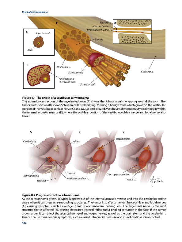
This textbook page contains 2 illustrations showing the progression of a vestibular schwannoma (acoutic neuroma). The first illustration highlights how the schwannoma begins within the internal acoustic meatus. The second illustration contains three stages of growth as the schwannoma grows into the cerebellopontine angle.
1. Clancy J. (2011) The Human Body: Close-Up. Firefly Books, Richmond Hill. 320 pp.
2.Kandel ER, Schwartz JH, Jessell TM. (2000) Principles of Neural Science, 4th edition. McGraw-Hill Medical, New York. 1414 pp.
3. Kiernan JA and Barr ML (2009) Barr’s the Human Nervous System: An Anatomical Viewpoint. Lippincott Williams & Wilkins, Philadelphia. 424 pp.
4. Lescanne E, Velut S, Lefrancq T, Destrieux C. (2002) The internal acoustic meatus and its meningeal layers: a microanatomical study. J Neurosurg 97(5): 1191-7.
5. Rengachary SS and Wilkins RH. (1994) Principles of Neurosurgery. Wolfe, London. 575 pp.
6. Standring S. (2005) Gray’s Anatomy: The Anatomical Basis of Clinical Practice, 39th edition. Elsevier-Churchill-Livingstone, Philadelphia. 1627 pp.
7. Wilson-Pauwels L, Akesson EJ, Stewart PA, Spacy SD. (2002) Cranial Nerves in Health and Disease, 2nd edition. BC Decker Inc, Hamilton. 245 pp.
8. Yamakami I, Nakamura T, Ono J, Yamaura A. (1999) Recovery of hearing after removal of a large jugular foramen schwannoma: report of two cases. Surg Neurol 51: 60–65.


